Acropora ID mini-class

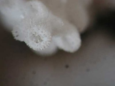
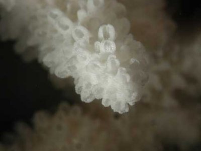
Here is one I would like to identify. A whole colony was brought to a PSAS meeting and this image is of my fragment of that colony. It has encrusted on rock at the very back of the tank and cannot be readily removed to be photographed so the image here is poor. The fragment was mounted on it's side and instead of starting new growth from it's upper side coralites(what I expected), the existing axial coralites are bending up as they grow.

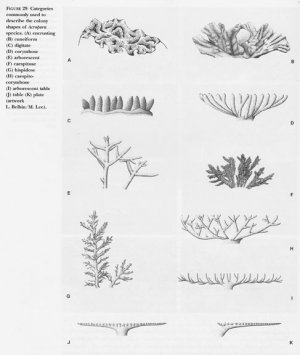
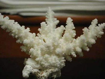
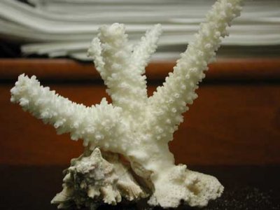
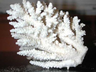
As I recall, there was a lot of anastomosing branches on the colony, especially in the lower, older, regions. The tips of the entire colony rose up to an even level like a tabletop, but this may be because they had reached the top of the water in the tank, I don't know. A form description that best applies - I would say - is corymbose. The anastomosing branches were generally in the lower, horizontal region and they terminate in short, upright branchlets. My fragment picured here is just an upright branchlet (mounted on it's side).

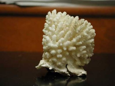
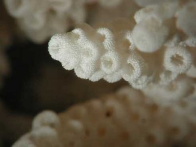
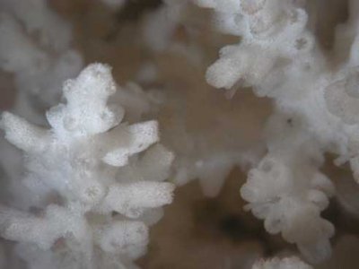
An image of a skeletal tip follows this post. I apologize for attaching photos rather than links, but the RC upload to gallery function is currently broken.

*edit* The upload image fuction also appears to be broken to attach images directly to a post. Hmmm.... I'll have to post back with links. Sorry.
 
JB - looks like you have a fairly regular corallite distribution...should be a good choice (in contrast to mine!)

I would call yours caespitose to corymbose. Hispidose corals have fine branches off the main branch. A classic example would be something like A. echinata.

howard - looking forward to seeing your photo.

I will post the axial information in a while.
 
EricHugo said:
JB - looks like you have a fairly regular corallite distribution...should be a good choice (in contrast to mine!)

I tried to pick something that the distinctions would be easier to notice.

EricHugo said:
I would call yours caespitose to corymbose. Hispidose corals have fine branches off the main branch. A classic example would be something like A. echinata.

Caespitose I was thinking about as well, but your description stated that the branches interlock. I don't see any of the branches fused together.

Actually I find this first part, IDing the type of growth form, for some reason very difficult for me.

Like why would you think that the coral I chose has a corymbose growth form. Wouldn't that kind of growth be similar to a classic table such as A. cytherea and A. hyacinthus?
 
JB it is hard, you're right. Its a subjective distinction in many cases, with overlap and variation made all the more difficult by the size of the colony. I would have said something very different from the first photo to the second, but as the colony developes, you see different characters. So, here is an image from Wallace 1999 that shows "idealized" growth forms. Would that it was this easy.

So, here is where familiarity with variation comes in. The definition is a "loose" one. Degrees of connectivity... well, look at the fusion of a common base brought by extreme anastomoses. But, tables like A. hyacinthus or A. cytheria are not corymbose. A. millepora is corymbose. But, when young, A. hyacinthus and A. cytheria might look corymbose, and others in the hyacinthus group even have corymbose characters as well-developed colonies.

Mine is actually a cross between caespitose, caespito-corymbose, and digitate. I have a feeling the digitate nature might become less if the colony were larger.

Your case exemplifies the change over relatively small time frames and sizes how deceptive ID of a small colony growth form can be. I have received numerous small colonies over the years that were sold as "tables" but really weren't.

Fortunately, this is just one step in the "narrowing down" process, and it is either helps, or it doesn't. For most available in stores, it is probably mostly unhelpful. For those that have grown quite a bit in tanks, maybe a bit more helpful, depending on how determinate the growth form for that species is across environmental gradients.
 
idealized growth forms

from Wallace, CC. 1999. Staghorn Corals of the World. CSIRO, Queensland, p. 52.
 

Attachments

  • growth-forms.jpg
    growth-forms.jpg
    39.2 KB · Views: 1
ok piercho - looks good.

So, other corals besides Acropora may have dimorphic polyps/corallites, but the characteristic is pronounced in Acropora. The axial corallites make up the axis of a branch, and the branch can sort of be considered as one long formation of the axial, with the solid part of the branch being the outer wall of the axial, with radial corallites budded off from below the growing axial. Sometimes, as in my photo, it can be difficult to tell which are really axials. Same is true for some hispidose colonies. Also, if growth slows or ceases, axial length by be reduced or even halted. Sometimes, the axial is very long and tubular...sometimes it is short and berry-like.

Contributions of branch thickness may be formed mainly from axials, radials, or both. But the wall of the axial corallites is formed from synapticular rings of varying numbers. If you look down directly into the axial corallite, you will see the septa. In some cases, they are very immersed deep in the corallite, while in others they are more apparent. There will be either six or twelve septa, arranged in one or two cycles. By cycles, I mean that there may be primary septa that are typically larger, thicker, and reach farther towards the center of the corallite. Secondary series are usually smaller, thinner, and do not extend as far towards the center. If you look at enough axials, you might find variation, with sometimes only 4 or 5 of the 6 secondary septa showing any development at all, so that it appears at first look as though there is not a full set. In other cases, primary and secondary are almost indistinguishable. In still others, there are only primary septa. You may also find that one set of primary septa are larger than the rest of them. These are called the directive septa.

I will take some photos later of different axials, including my specimen. These characters of your axial corallites should be written down. Also important is to take measurements. You will need to known the outer diameter of the axial corallite (outer wall to outer wall) and the inner diameter (from inner wall to inner wall - basically across the void into which the septa penetrate). You should use a millimeter scale or calipers to determine these measurements. You should also take a measurement of several axials and take the mean of them. There can be considerable variation in them across a colony. Alternately, you could measure the smallest fully developed axial and the largest, and this would be the range of diameters.

All of this information will be used in determining the species. You may see already how impossible it is to determine this information in a living coral where tissue covers these minute skeletal characteristics.
 
Thanks for that image from Wallace, pictures are worth a thousand words. OK I see why A. millepora, not A. hyacinthus, would be considered corymbose. I've seen that kind of anastomoses on some of my larger A. millepora colonies.

Looking at that table, my coral most resembles a caespitose-corymbose growth form, letter H on the picture.
 
I would agree with you.

So, I did not take my camera to lab today, so I am trying to get a good photo by hand of my Acropora. I am also having to use a ruler, which you may have already discovered is not an easy thing to do.

So, Until tomorrow when I use the scope, my coral has two cycles of septa, no clear directive septa, and the secondary series is highly irregular and imperfect. In many axials, I only see one or two secondary septa, and they do not show any real consistency as to how far towards the center they go. The primary septa extend about 50-60% of the radius. The outer corallite diameter is about 1.8-2.0mm and the inner diameter appears to be about 0.5-0.75mm, although this is at best an estimate using a ruler. Here is what it looks like.
 

Attachments

  • untitled-1.jpg
    untitled-1.jpg
    5.1 KB · Views: 3
Yokes, dude, this is almost comical with a 20X loupe. You made me go chop off a new 3-axial piece to get this done.

# Septa: 12
# Cycles: 2
Notes on septa: the secondary septa are inconsistently apparent on some axial coralites. Rarely you can count all 12 septa, sometimes just >6 & <12. My primary septa don't extend nearly as far out into the center as the ones you show do, and are not always distinct from the secondaries.
Outer diameter: ~2.4 mm, until I can get a better scale. We are talking about all the way across the end of the tip here, right?
Inner diameter: 0.8 mm, estimated. Roughly 1/3 of the outer diameter, by eye.

Here was something I found interesting: the new bit I chose to chop off had a anastomized (use?) branch joining it. I expected to have to clip it twice to free it. When I made the first clip, it popped right off the branch growing into it - which had a blunt cylindrical end covered with flesh. The clipped fragment has a 1/4" deep socket where the anastomized branch grew into it which was bare (no apparent flesh) and slick. I suppose this would allow these pieces to flex on some axis - rather than break - if mechanically stressed.
 
You guys still with me or am I just typing for nothing? If so, let me know, because I have lots of other things I could be doing....

here are some more images of axials and the corals they came from.
 
Back
Top